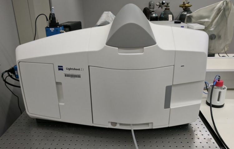
Zeiss Z.1 Lightsheet
Description
Thanks to the NIH , Head PI Bill Smith, and Co-PIs Denise Montell, Anthony De Tomaso, Craig Montell, Joel Rothman, Kenneth Kosik, Samir Mitragotri, and Otger Campas.
This lightsheet microscope is fully equipped and can be used for fast imaging of samples with a lot of 3-dimensional depth. Most commonly used for imaging embryo development, and other multicellular organsims live or fixed. Excellent environmental chamber controls for temperature, immersion media, and CO2. Instead of imaging on a slide, the sample is embedded in a column of agarose.
There are 6 laser excitation lines (405, 445, 488, 515, 561, and 638 nm). Dual 1900x1900 pixel cooled scientific CMOS cameras for two-color simultaneous imaging. Dual filter sets for DAPI-GFP, CFP-YFP, GFP-mCherry, GFP-DRAQ5, or CY3-DRAQ5. The three objectives are a 40x/1.0, 20x/1.0, and 5x/0.16. All three are designed for your sample to be immersed in media with index of refraction = 1.33 (water), and the 5x can be adapted for CLARITY (index of refraction = 1.45).
For now the facility director (Ben Lopez) will handle sample chamber and objective lens preparations, so each reservation will need to include an indication of live or fixed media, and choice of objective lens.