Nanolive 3D Cell Explorer
Description
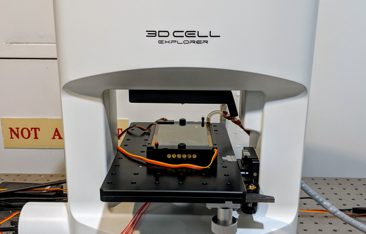
The Nanolive 3D Cell Explorer is a novel new instrument that uses holographic tomography to generate a three-dimensional image of the sample with contrast determined by refractive index. It works primarily with adherent cell cultures where the instrument makes many subcellular structures immediately visible label-free. The cell membrane, nuclear material, nuclear envelope, condensed chromsomes, mitochondria, and lipid droplets are all clearly identifiable. A 30 micrometer stack of images can be collected every 2 seconds. It uses a low powered green laser that does essentially no photodamage; that combined with the included environmental chamber can keep cells alive in a healthy state for days at a time allowing incredibly long timelapses. The system also includes 4-channel fluorescent imaging with blue, green, red, and far-red filters.
While adherent cell cultures are the most common subject for imaging on this device. Bacteria and other microbes can be visualized and tracked. On the larger end of the spectrum, C. elegans organisms and their internal structure are clearly visualized.
See this link for example videos: https://nanolive.ch/applications/