Instrumentation

Leica SP8 image

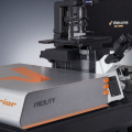
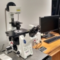
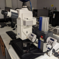
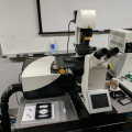
The new fast resonant scanning Leica SP8 confocal microscope is a high-tech powerhouse. The resonant scanning unit allows confocal images at 28 frames per second full-frame, and faster for smaller images. It is equipped with a cryo-incubator stage allowing stable temperature control from -2°C to 50°C enabling live real-time long time-lapse imaging. The recent upgrade has added FLIM (fluorescence lifetime imaging microscopy) capabilities!

This amazing new microscope uses Stimulated Emission Depletion to capture super resolution images with as fine of resolution as 30-50 nm, which is a huge improvement compared to the typical resolution limitation of 200-300 nm with a normal confocal microscope. It is compatible with fixed and live samples and sample prep is similar to confocal. Certain fluorophores work better for STED than others, so be sure to consult with the facility director for help planning your sample preparation. Also works great as a normal confocal microscope, especially for low signal imaging!

JEOL 1230 TEM

The JEOL JEM-1230 is a high performance, high contrast, 40-120kV transmission electron microscope with excellent imaging capabilities suitable for biological and materials science applications.

Nanolive 3D Cell Explorer

The Nanolive 3D Cell Explorer is a novel new instrument that uses holographic tomography to generate a three-dimensional image of the sample with contrast determined by refractive index. It works primarily with adherent cell cultures where the instrument makes many subcellular structures immediately visible label-free.  A 30 micrometer stack of images can be collected every 2 seconds. It can keep cells alive in a healthy state for days at a time allowing incredibly long timelapses.

Zeiss Lightsheet microscope

This lightsheet microscope is fully equipped and can be used for fast imaging of samples with a lot of 3-dimensional depth. Most commonly used for imaging embryo development, and other multicellular organsims live or fixed. Excellent environmental chamber controls for temperature, immersion media, and CO2. Instead of imaging on a slide, the sample is embedded in a column of agarose.

Spectral Confocal

This system is an inverted Olympus IX81 confocal microscope equipped with 6 laser lines for excitation, spectral detectors and environmental control. 

An upright microscope with DIC optics, an alternate high NA darkfield condenser and fluorescence.

Olympus BX60

Olympus BX60 Microscope an Upright Compound Microscope with DIC optics, fluorescence and Olympus DP73 color camera

IX81 inverted microscope

This Olympus inverted compound microscope is equipped with high quality objectives, fluorescence and a Qimaging scientific CMOS camera allowing high frame rates even in low light conditions. The filters, objectives, and z-position are all motorized for automated image collection.

Zeiss Axiovert Image

Inverted motorized compound microscope with phase optics, fluorescence, and CCD camera.

The Zeiss Axio Zoom Stereoscope has transmitted and reflected brightfield, fluorescence, and a motorized stage. Great for lower resolution imaging of large samples or entire tissue slices or slides. Big thank you to the Denise Montell Lab for letting us offer this to users while the facility houses it for them!

Olympus SZX12 Stereomicroscope

The Olympus SZX12 stereomicroscope is used for tissue preparation and documentation of samples at lower magnifications.

Imaris Software

This computer workstation runs current Imaris software with the Imaris Measurement Pro, Filament Tracer, Cell, Colocalization, Track, Vantage and XT modules. It also has Zeiss Zen, Leica LASX, and Huygens deconvolution software.

Cryostat

This Leica Cryostat CM1850 is designed to cut thin sections of frozen material.

The Leica VT1000S vibratome is designed to cut sections (50 microns and up) of fresh material or fixed material. Usually the material is embedded in a high-percentage agarose gel. The vibratome is free to use, but you still need to be trained on it and schedule time on the scheduling site. Please bring your own consumables (blades, superglue, buffer solution, etc.).